Phylum Platyhelminthes – Webquest Notes
Rather than me going through the notes and boring you, today you will be using this website to go over the notes, videos, and pictures at your own speed. You will gather notes through answering questions which gives you examples of questions I could ask on the simple invertebrates test while adapting my notes to fully address the question. Feel free to use other materials/videos to explore these phylum and help clarify if you need. If you would like me to check on any of your answers/diagrams, please let me know!
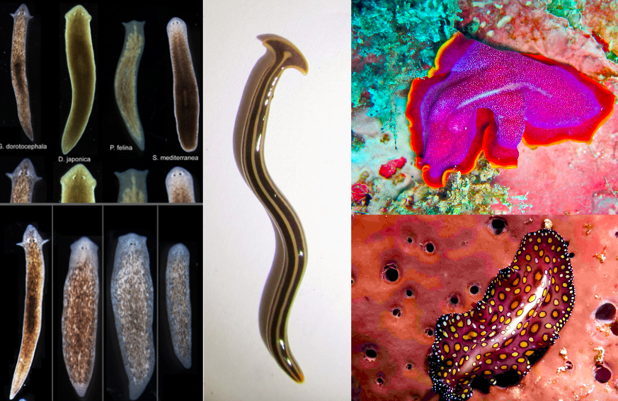
Platyhelminthes -> “Flat” “Worm”
- Appeared ~540 – 500 Million Years ago
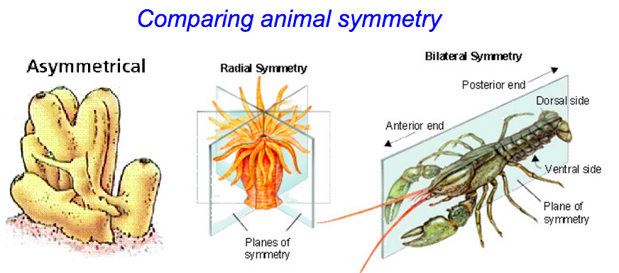
- First animals with bilateral symmetry and cephalization (sense organs to the “front” – development of brain)
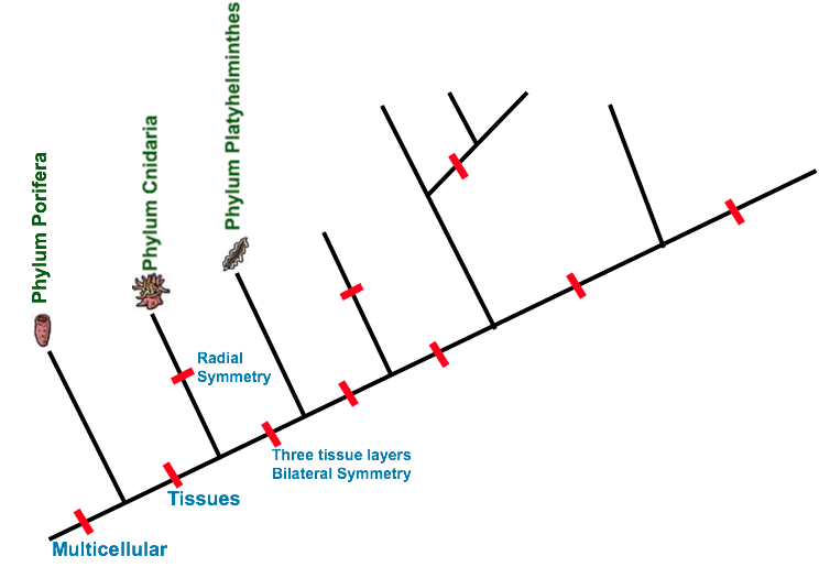
A: Evolution of Body Plan
1. Three tissue layers – Endoderm, Mesoderm (muscle, organs), and ectoderm. They are triploblastic.
- Flatworms (like sponges/cnidarians) are acoelomates. This means they have no body cavity.
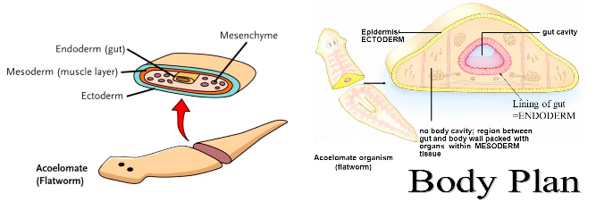
Looking back: Tissue development continues (Porifera – no layers, cnidarians – 2 layers filled with jelly-like mesoglea), flatworms – 3 layers)
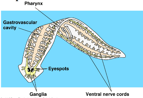
2. Cephalization – Development and concentration of nerve tissue at one end of the body. Accessory organs for hearing, seeing, taste developed in this area.
- First animals to evolve a nervous system
- Eyespots – Accessory organs used to detect intensity of light
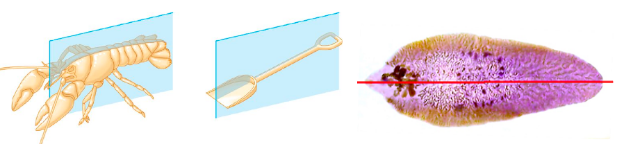
3. Bilateral symmetry – The body plan can be divided in half making a right and left side that are mirrors of each other.
B. Feeding and Excretion
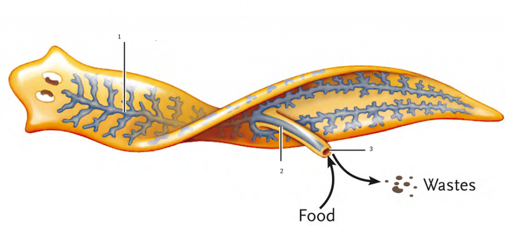
- Two-way digestive system – Food is ingested and waste is expelled through the pharynx.
- No mouth – a pharynx is present on the underside of the animal that goes directly into the gastrovascular cavity.
Two Methods of Feeding:
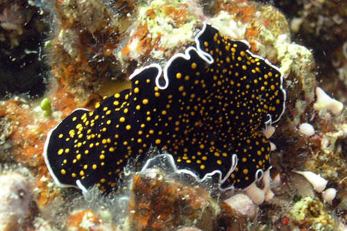
- Free-living
- Carnivores that eat tiny aquatic animals or detritivores scavenging from dead animals.
- The gastrovascular cavity forms an “intestine” that has many branches all throughout the entire length of the worm.
- Parasites
- Feed on blood, tissue fluid, or pieces of cells within host.
- Typically have adaptations such as suckers/hooks to attach to host
- Digestive system is much simpler that free-living flatworms

Excretion
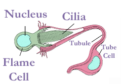
- Simplest animals to have excretory system
- System of simple tubles running throughout the body that open to the outside via pores.
- cells in the tubules are called flame cells because they have a cluster of cilia (hair) that looks like a flickering flame.
C. Respiration and Circulation
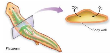
- Just as sponges and cnidarians – flatworms have no internal transport network.
- Gas exchange occurs through diffusion (O2 in, CO2 out) through the skin.
D. Reproduction
1. Free-living flatworms:
- Hermaphrodites that can reproduce sexually or asexually.Sexually
- 2 worms pair up and both worms receive sperm (internal fertilization of both or “penis fence” winner)
- Eggs are laid in clusters
Asexually:
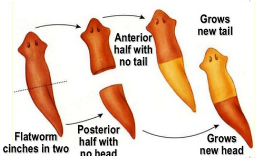
- Fission/Fragmentation – Flatworms can regenerate into new clones of the parent after purposeful splitting or damage. (Fission – Purposeful, Fragmentation – Accidental)
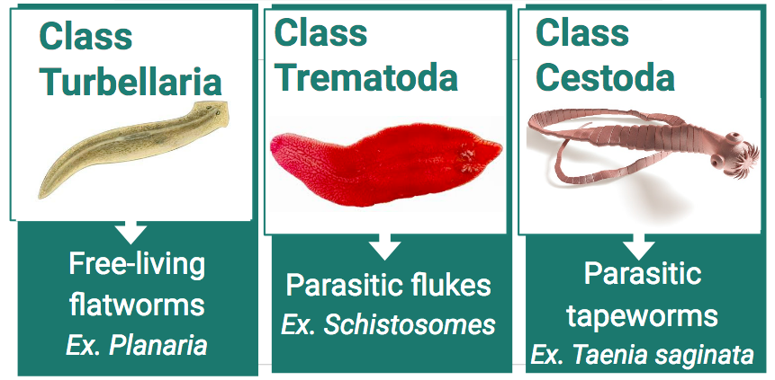
2. Parasitic Flatworms
Typically have very complex life cycles involving one or more host. Please choose one of the following life cycles to learn about and be ready to explain.
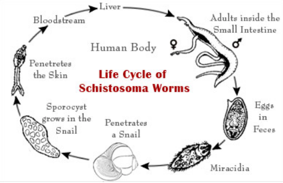
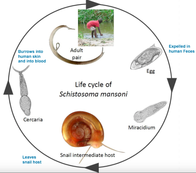
A. Schistosomes (Group of blood flukes) Causes schistosomiasis in humans
Uses snails and humans as hosts to complete its life cycle.
- Eggs are laid in human intestine until their is a rupture.
- Blood and eggs leak into the intestine and enter human feces.
- Feces ends up in streams/fields where the eggs hatch into free swimming larvae.
- Larvae enters an intermediate host (snail). Larvae grows inside snail and eventually leaves.
- Worms travel through the circulatory system – live in hear, lungs, or intestines.
- Free-swimming worms bore into human skin and enter blood
Schistosomiasis can result in death, kidney failure, growth problems, etc.
-
-
- a high temperature (fever) above 38C.
- an itchy, red, blotchy and raised rash.
- a cough.
- diarrhoea. (bloody stool)
- muscle and joint pain.
- abdominal pain.
- a general sense of feeling unwell.Symptoms of schistosomiasis include:
-
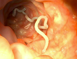
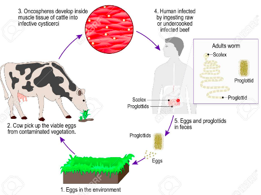
B. Tapeworms – Taenia saginata (beef tapeworm) and T. solium (pork tapeworm)
Uses humans and cows (T. saginata) or pigs (T. solium) as hosts to complete its life cycle.
- Eggs or proglottids (asexually created with male/female parts that break off) are passed from humans with feces.
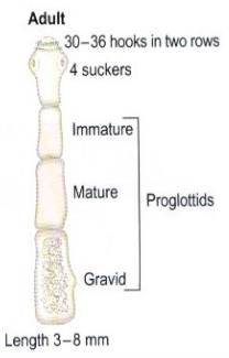
2. Cows/pigs are infected by eggs through contaminated vegetation.
- Eggs hatch in animal and burrow through intestine into muscle where they develop into cysticerci (can survive many years)
4.Humans ingest raw or undercooked meat with cysticerci.
5. Cysticerci develops into adult tapeworm and attaches to the intestine with a scolex (hooks/suckers)
Length of adult worms is usually 5 m or less for T. saginata (however it may reach up to 25 m) and 2 to 7 m for T. solium.
- The adults produce proglottids which mature and detach from the tapeworm and passed with feces (approximately 6 per day)
E. Major Groups
Comments by shaun pletsch