This section is mostly focused on the exciting world of memorization. There are a lot of pieces and important structures in the heart and circulatory system and this section is focusing on building the terminology and the “map” of our system.
The Heart
Four-chambered with double-loop system.
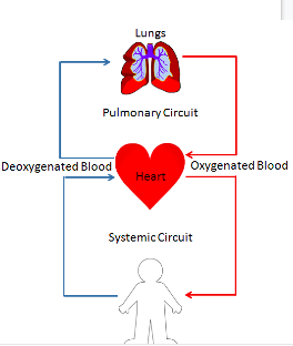
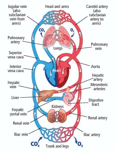
- Double-loop circulatory system – one “loop” that goes to the lungs and back, one that goes from heart to body and back. (Although as we will learn it is more a “figure 8” path due to mirrored sides)
- Pulmonary Circuit – Pumps blood from the right side of the heart to the lungs.
- Systemic Circuit – Pumps blood from the left side of the heart to the body
The heart is a muscular organ located in the chest.
- Walls are composed of cardiac muscle held together by connective tissue.
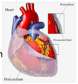
- A tough membranous sac surrounds the heart. This is called the pericardium. The pericardium is filled with fluid which reduces friction as the heart beats.
Basic Structure:
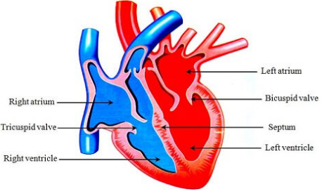
The heart has two sides separated by a muscular septum. The pumping action is synchronized; muscle contractions on the right side mirror those on the left side.
1. Two-thin-walled atria (singular: atrium) are the receiving chambers.
- Right Atrium – Receives deoxygenated blood from the body.
- Left atrium – Receives oxygenated blood from the lungs
2. Two-thick walled, muscular ventricles pump blood out to distant tissues.
Think: Why could the left ventricle be more muscular than the right ventricle?
3.The Valves (or why blood doesn’t flow backwards):
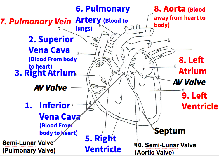
- Semi-lunar Valves – Found at the beginning of arteries. At the base of the aorta it is called the aortic valve. At the base of the pulmonary artery it is called the pulmonary valve. This shut after ventricle contraction since the blood pressure is lowered in the arteries after contraction.
- Atrioventicular (AV) Valves – Separate atria from ventricles. Chordae tendineae are tiny tendons that attach the valve flaps to the interior of the ventricle.This ensures blood does not flow backwards during ventricular contraction (like a one way door held closed by these tendons)
Tracing Blood Through the Heart
- Deoxygenated blood enters the right atrium from the body through the vena cava.
- Atria contract, pushing deoxygenated blood through the right atrioventricular valve and into the right ventricle.
- Ventricles contract, pushing blood through the right semi-lunar valve (pulmonary valve) and into the pulmonary artery towards the lungs.
- Gas exchange occurs at the lungs, oxygenating the blood and removing CO2.
- Oxygenated blood enters the left atrium through pulmonary veins.
- Atria contract, pushing the oxygenated blood through the left atrioventricular valve and into the left ventricle.
- Ventricles contract, pushing blood through the left semi-lunar valve (aortic valve) and into the aorta out towards the body.
- Oxygenated blood moves around through the body’s arteries where gas exchange occurs in the capillaries.
- Deoxygenated blood leaves the capillaries towards the heart.
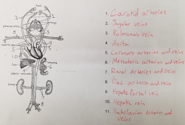
Major Vessels in the Circulatory System
Below is the link to the padlet created by the class. Use the quick review table handed in class for basic locations.
https://padlet.com/mrpletsch/bio12vessels
Password: kill
Heart Control
The heart possesses its own specialized conduction system and can beat independently even after being separated from its nerves (that means the heart can beat after it is removed from the body until it dehydrates or loses ATP)
- The extrinsic (arising external to the heart) nerves coming from the nervous system serve to modify and control the beating established by the heart, not just force it to beat.
- The sympathetic nervous system seeks to increase heart rate in times of stress/need. “Fight or Flight”
- The parasympathetic nervous system seeks to slow heart rate down in times of rest/digestion. “Feed and Breed”
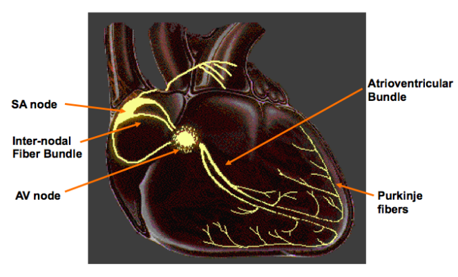
The heart has two areas of specialized tissue both located in the right atrium:
1. The Sino-Atrial (SA) node – Initiates contraction of the heart (average = 72 times/minute). Called the pacemaker.
2. The AV node – Causes ventricular contraction after ventricles are “full.” Uses purkinje fibres (set of nerves that send impulses, coordinating ventricular contraction).
- The AV node serves as a conductor, passing nerve impulses via two large nerve fibers, called Purkinje fibers.
- The Purkinje fibers run along the septum carrying impulses from the AV node to the bottom tip of the heart.
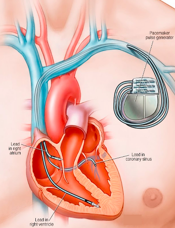
People with irregular heartbeats may require an artificial pacemaker to stimulate the SA node.
- The little device is connected so that it can stimulate the SA node with electric current
- This causes initiation of the cardiac cycle (basically just kick starts the cycle when the SA node has troubles)
Heart Rate and Cardiac Cycle
Cardiac Cycle – Complete series of events leading to one full heart contraction.
The double sound of a heart beat is caused by the closing of two different sets of valves: (AV valves/Semi-lunar Valves)
- Lub – Closing of AV valves after blood enters ventricles
- Dub – Closing of Semi-lunar valves after blood leaves ventricles
ECGs and Introduction to Irregularities
We can look at these nerve impulses in individuals to determine abnormalities in the cardiac cycle and diagnosis certain heart conditions.
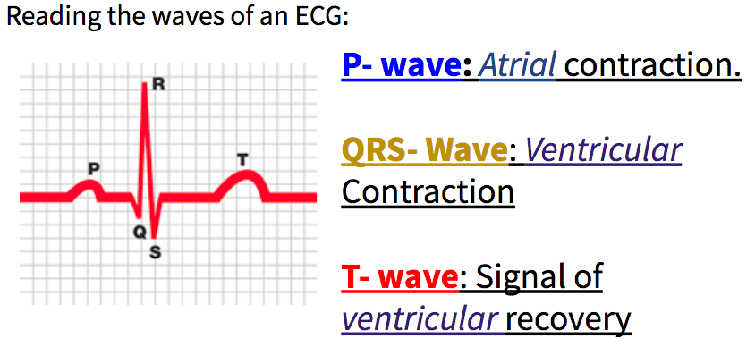
- The electrical impulses are displayed on a graph called an electrocardiogram (ECG).
- Changes in electrical current reveal normal or abnormal events of the cardiac cycle.
On average humans complete the cardiac cycle 72 times a minute.
Identifying basic irregularities on a ECG:
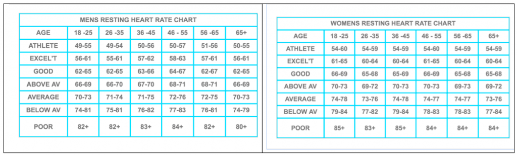
Tachycardia = heart rate of greater than 100 beats per minute.
Bradycardia = Heart rate lower than 60 bpm (although many athletes can achieve this without it being considered an issue)
Artificial pacemaker = Pacemaker will give an electric current to start cardiac cycle. This can be observed on an ECG before the wave complex begins
Atrial Flutter = Type of tachycardia, but the atria contraction are irregular and out sync with ventricles.
Sinoatrial arrest = Failure of heart contraction due to issue with SA node (greater than 2 seconds).
Reading Irregulary ECG practice sheet – Answer key to the four cases (Click here!)
Quiz Expectations
Circulatory System Quiz 2: What to know
1. Heart Structure – Identify the following on a diagram/example.
| Atria | Ventricles | AV Valves |
| Semi-lunar Valves | Aorta | Pulmonary Artery |
| Pulmonary Vein | Inferior Vena Cava | Superior Vena Cava |
| Septum | Choridae Tendinae |
- Brief description (i.e. atria thin walled receiving chambers, ventricles thick, muscular walled to pump to pulmonary or systemic circuit).
- Identify which of the above carries deoxygenated/oxygenated blood (i.e. right atrium/right ventricle get deoxygenated blood from the body)
- Track blood through the structures of the heart and around the body
2. Major Vessels – Identify the following on a diagram or explain its rough location (i.e. hepatic portal vein – runs from intestine to liver).
| Aorta | Superior/Inferior Vena Cava | Mesenteric Arteries and Veins |
| Hepatic Portal Vein | Hepatic Vein | Renal Arteries and Veins |
| Iliac Arteries and Veins | Subclavian Arteries and Veins | Carotid Arteries |
| Jugular Veins | Coronary Arteries and Veins | Pulmonary Artery and Veins |
3. Heart Control – The notes from today – nothing outside of this package. Differentiate between when the parasympathetic vs. sympathetic nervous system would influence heart rate.
4. Reading an ECG – Label/discuss P, QRS, and T waves. Identify a common irregularity in an ECG.
Comments by shaun pletsch