Respiration – Defined
1. Structures of the Respiratory System
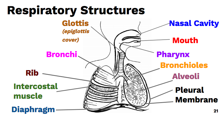
- Nasal Cavity – The nasal cavity is the inside of your nose.
- Lined with a mucous membrane that helps keep your nose moist.
- Cilia – Trap dust, debris
- Warms and moisten air
- Mouth – Another pathway for air into the respiratory system.
- Pharynx – a common pathway for food and air. Nasal cavity and mouth meet here.
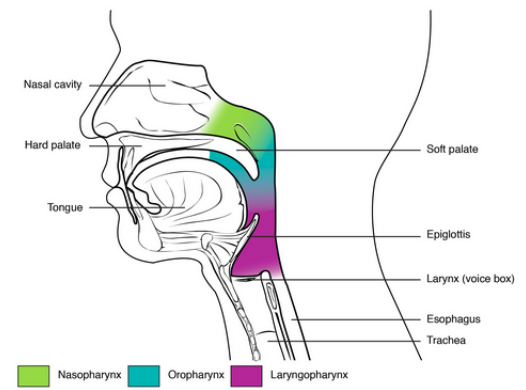
- Glottis – Opening to the Larynx (voice box). When we breath the glottis is open, when we swallow the epiglottis covers the glottis is prevent food entering the larynx.
- Epiglottis – Flap that covers the glottis. Prevents food from entering the lungs.
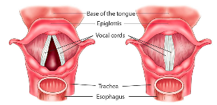
- Larynx – Triangular, cartilaginous box that contains the vocal cords and regulates air entry into the lungs.
- Vocal cords – Vibrate as air moves past, producing sound.

- Trachea – “Windpipe.” The sturdy pathway for air to the lungs. Like the nasal passage, it is covered in cilia and a mucous membrane.
- Held open by cartilaginous rings.
- Cilia beat upward moving mucus and any particles away from the lungs. Smoking destroys cilia!
- Bronchi/Bronchioles – The trachea branches into two bronchi which are the pathway into the lungs. The bronchi branch into even smaller pathways called bronchioles which get narrower and thinner walled as they go onward (bronchioles are not supported by cartilage rings)
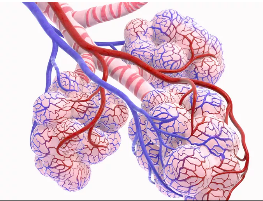
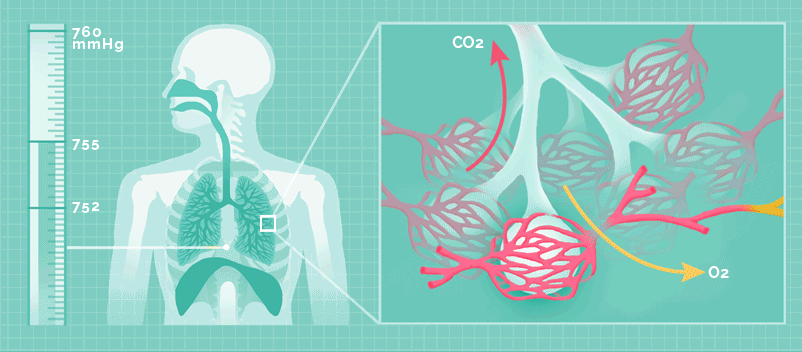
- Alveoli – Sacs at the end of bronchioles where gas exchange occurs.
- One cell thick for rapid gas exchange with blood.
- Expand and stretch as air enters.
- ~300 million alveloi per lung (150 m2 of alveolar area)
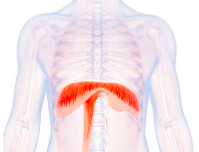
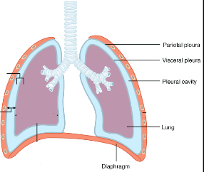
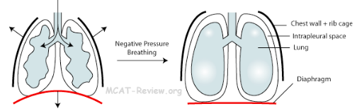
- Diaphragm – Thick, dome shaped muscle at the bottom of the thoracic (chest) cavity. Separates abdomen from chest. Attached to ribs, sternum, and vertebral column.
- Intercostal muscles – Muscles between ribs that helps seal the chest cavity and expand the chest cavity during breathing.
- Pleural Membrane – Membranes that allow for an air-tight seal around the lungs.
- One pleural membrane lines the chest walls.
- Inner membrane lines the lung. In between is fluid.
2. Mechanism of Breathing
How does air enter and exit the lungs?
The intercostal muscles, diaphragm, and the pleural cavities allow us to pull air in and push air out of our lungs.
- These structures create negative pressure inside our lungs.
- Negative Pressure: Air pressure less than the surrounding air (760 mm Hg)
Think: How can we create negative pressure in our lungs?
This negative pressure is created by increasing the volume inside the thoracic cavity.
Air will naturally move in to fill this extra volume created, moving into the lungs and balancing the pressure inside and outside the lungs.
So how can we increase the volume of the thoracic cavity?
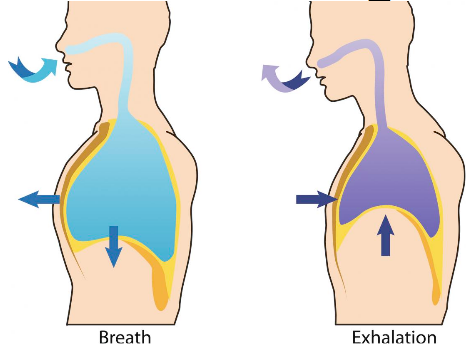
Inhalation – Air Moving in
- Intercostal Muscles – contract and pull the ribs up and out.
- Diaphragm – Contracts and pulls downward expanding the thoracic cavity.
Pressure inside lungs < than atmospheric pressure. Air moves in.
Exhalation/Expiration – Air moving out
- Intercostal Muscles – Relax. Ribs move in and down. If you push out air even harder, you force the muscles to push the ribs inward past relaxation.
- Diaphragm – Relaxes. Return to curved shape.
Pressure inside lungs >than atmospheric pressure. Air pushed out.
Comments by shaun pletsch