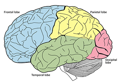
Exploring the lobes of cerebrum:
Phineas Gage – A railroad construction foreman that had an accident leading to extreme brain trauma. He survived, but his friends and family said he was unable to regulate emotions and was quick to anger. His personality changed completely. What part of the brain do you think was injured?
Injuries to the Occipital Lobe:
- Epilepsy: Some seizures occur in the occipital lobe, and occipital lobe damage increases vulnerability to seizures.
- Difficulties with movement: Even if you are still able to move, changes in depth perception and vision can lead to inappropriate movements and difficulty navigating the visual field.
- Difficulties perceiving colors, shape, dimension, and size.
- Difficulty recognizing familiar objects or faces.
- Hallucinations.
- Inability to detect that an object is moving.
Injuries to the Temporal Lobe:
- Various forms of aphasia, a disorder of speech and language.
- Impaired memory skills. Common problems include difficulty recognizing people, faces, or objects; poor long-term memory; disturbances in autobiographical memory, and poor auditory memory.
- Changes in self-image and self-perception: Because the temporal lobe houses many of our memories, disruptions in or loss of autobiographical memory can produce personality changes, as well as changes in a person’s sense of self.
- Changes in executive function. Some people with temporal lobe damage struggle to plan or coordinate their actions.
- Issues with smell – unable to identify.
Injuries to the Parietal Lobe:
- Right parietal lobe damage can impede your ability to care for your body because it undermines your ability to notice or care for at least one side of the body.
- Struggles with writing, arithmetic, math, and the ability to perceive objects.
- Impedes motor skills and visual attention. Do not know left from right and cannot integrate visual cues with movement.
- Unable to perceive stimuli like pain and pressure. Or incorrectly perceiving this stimuli.
Don’t forget to review your brain puzzle for the quiz/test!
Part 2: Neurons
Neuron – basic working unit of the brain, a specialized and electrically excitable cell designed to transmit information to other nerve cells.
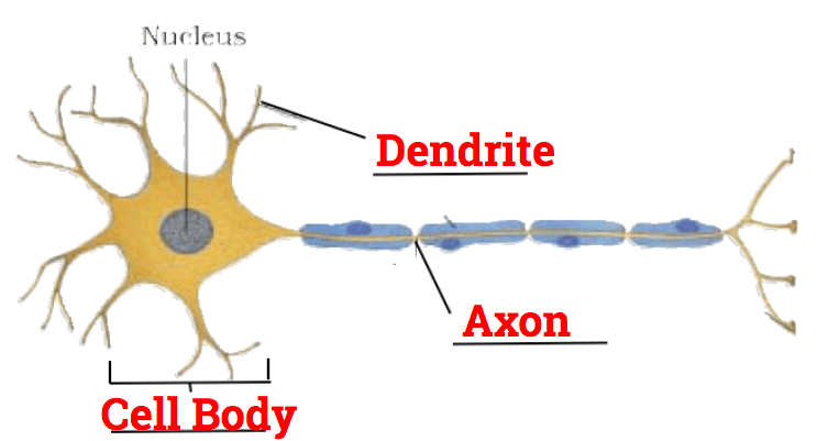
Three major parts of a neuron:
- Dendrite – Conducts impulses toward cell body. (Receives input)
- Axon – Conducts impulses away from cell body.
- Cell Body – Contains nucleus and maintains cell.
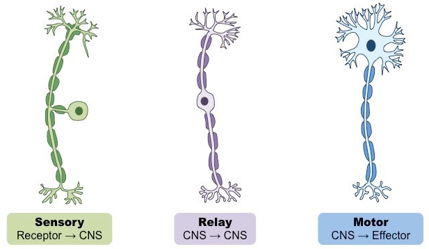
Types of neurons:
Sensory Neuron – convert external stimuli from the environment into internal electrical impulses. (Touch, smell, pain, etc.)
- Long dendrites that extend from receptors to the CNS.
- Relatively short axon that enters the CNS
Relay/interneuron – Connect sensory neurons to motor neurons within the CNS.
- Short dendrite
- Axon can be long or short
- Cell bodies of motor and interneurons create grey matter.
Motor Neuron – Conduct impulses from CNS to an effector (i.e. muscle)
- Short dendrite
- Long axon
- Dendrites/cell body are within CNS – axons are outside.
- Cell bodies of motor and interneurons create grey matter.

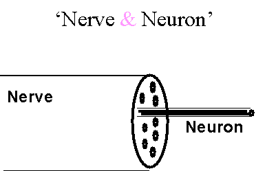
So is a neuron a nerve?
No!
- A neuron is a single cell of the nervous system.
- When combined with many other neurons, attached to a blood supply, and coated with a protective sheath, we have a nerve.
- It’s like thinking of a single muscle cell compared to the entire muscle.
Myelination and Nerves
- Nerves can contain both dendrites of sensory neurons and axons of motor neurons.
- Can have hundreds of “fibres”
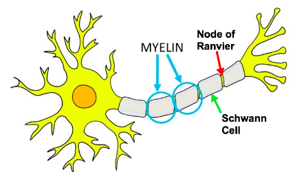
- These are protected by a fatty tissue called a myelin sheath.
- Myelin is composed of Schwann cells that wrap around the nerve fibre.
- Points between each Schwann cell is called a node of Ranvier.
Note: Individual neurons can be mylinated or unmylinated!
Think: What benefits could a myelin sheath give to a nerve?
- Insulation – prevent cross communication.
- Faster impulses
- Protection.
Reflex Arc
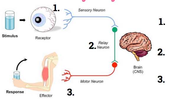
- Some sensory signals do not go to the brain before going to the effector.
- A receptor senses pain and the signal is sent through the sensory neuron to the CNS.
- It is transmitted to the motor neurons and out to the effector that causes a reflexive reaction.
- The brain is notified and process the signal after the reflex.
Benefit: This can protect you from serious, immediate damage
Comments by shaun pletsch