Rather than me going through the notes and boring you, today you will be using this website to go over the notes, videos, and pictures at your own speed. You will gather notes through answering questions which gives you examples of questions I could ask on the circulatory test while adapting my notes to fully address the question. Feel free to use other materials/videos to explore these concepts and help clarify if you need. If you would like me to check on any of your answers/diagrams, please let me know!
At this point you should have a road map of the circulatory system and understand the role of blood pressure. Now we are going to look more deeply at vessel structure and how exchange works in relation to this structure and through the concept of pressure!
A. Blood Vessels – Structure/Function
Blood Vessel: A tubular structure that carries blood through tissues and organs of the body (closed-circulatory system)
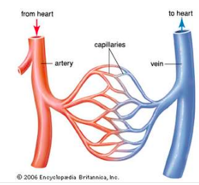
- Arteries: Carry blood away from the heart.
- Veins: Carry blood towards the heart.
- Capillaries: Network of blood vessels where exchange occurs.
Arteries:
Arteries typically carry oxygenated blood (systemic system).
- However, in the pulmonary system this is reversed – the pulmonary artery carries deoxygenated blood from the right ventricle to the lungs.
- Highest blood velocity.
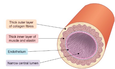
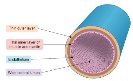
Structure:
- Have thick walls composed of distinct layers.
- The outer and inner layers are primarily connective tissue, while the thick middle layer is made up of muscles fibers and elastic tissues.
- This thick walled, elastic structure allows for expansion and changes in pressure.
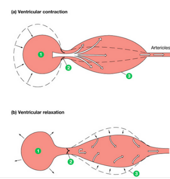
- Artery – Pulse/Pressure changes:
- ~70 ml of blood is pumped into the arteries each contraction.
- Arteries expand and stretch to facilitate the new blood flow. This change in diameter can be felt as our pulse
- Arteries relax and return to original shape before next contraction.
- Fun Fact! Some of the largest arteries have enough tissue mass that need to be supplied oxygen by smaller arteries!
Artery – Arterioles:
Blood from the arteries passes into smaller arteries, called arterioles.
- Arterioles’ diameter is controlled by the autonomic nervous system (not conscious). This can help maintain homeostasis – think medulla oblongata inhibiting or stimulating the vasomotor centre!
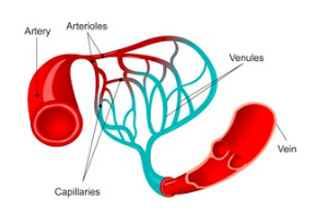
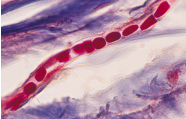
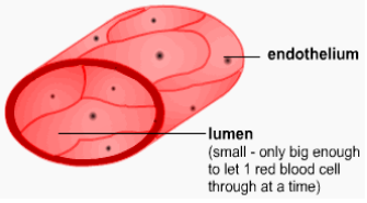
Capillaries:
The arterioles narrow and branch into single-cell wide capillaries.
- Capillaries are the location of fluid and gas exchange between blood and cells.
- No cell is more than two-cells away from a capillary!
- Slowest blood velocity due to branching paths, friction, and narrow size!
Structure:
- No muscle/elastic fibers
- Walls are a single layer thick.
- Capillaries are very fragile and can break through high blood pressure or an impact (like punching your friend…don’t try…)
- Blood filling the spaces between tissues causes bruising
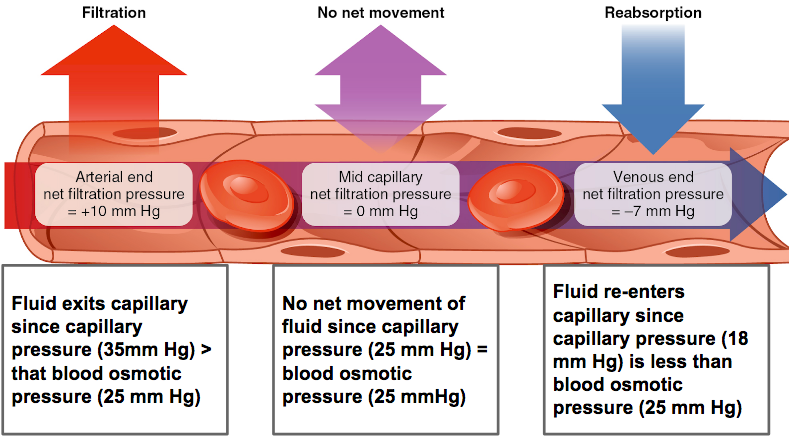
How does the exchange happen within capillaries?
Capillary – Exchange (Basic):
- Oxygenated blood enters the capillaries from arterioles.
- Hemoglobin in RBCs release oxygen due to temperature and pH change).
- The water, O2, and nutrients are forced through the thin capillary walls into the cells due to a higher pressure inside the arteriole side (35 mmHg).
- By the time the blood is on the venule (vein) side of the capillary, the pressure is now lower (18 mm Hg) and the blood is now hypertonic (less water than cells). Water moves in (osmosis) and CO2 and ammonia move in through diffusion.
Veins:
Veins typically carry deoxygenated blood (systemic system)
- However, in the pulmonary system this is reversed – the pulmonary veins carry oxygenated blood from the lungs to the left atrium.
- Higher blood velocity than capillaries, lower than arteries)
Structure:
- Thin walls with smooth muscle.
- Venules (from capillaries) merge into veins, which have a greater diameter.
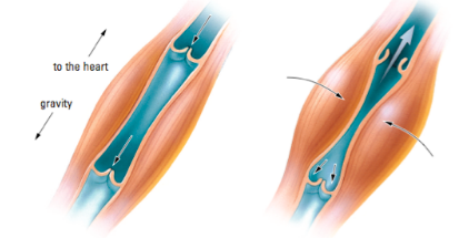
- Contain valves. Since the blood is pumping upward, valves prevent backflow in veins.
- Since the pressure in veins (~15 mmHg) is not enough to push blood against gravity towards the heart, skeletal muscles can contract and push the blood upward.
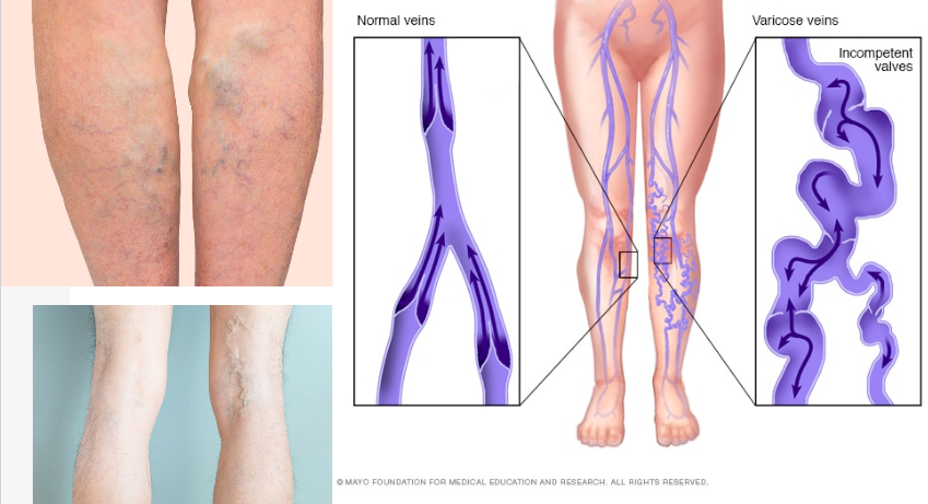
Improper valve function and varicose veins:
Varicose veins are usually caused by weak vein walls and valves.
If walls of the veins become stretched they can loose their elasticity, leading to leaky and weak valves.
If the valves don’t function properly, this can cause the blood to leak and flow backwards. If this happens, the blood collects in your veins, which become swollen and enlarged. We can these varicose veins.
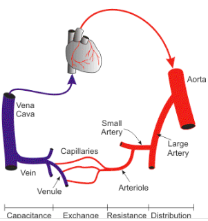
Blood Vessel – A Summary:
Heart -> Arteries -> Arterioles -> Capillaries -> Venules -> Veins -> Heart
- Arteries – Move blood away from heart
- Veins – Move blood toward the heart
- Capillaries – Site of gas/fluid exchange
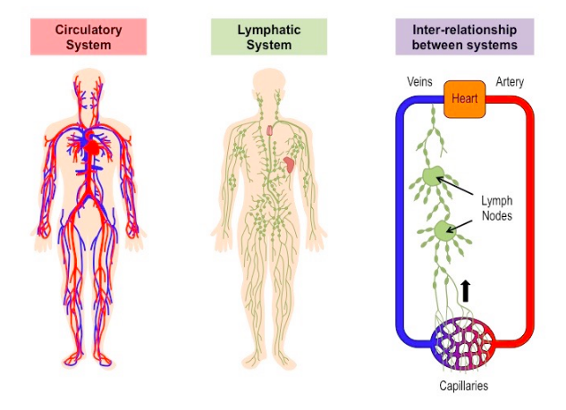
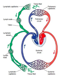
B. The Lymphatic System
The lymphatic system is not really a separate system of the body. It is considered a part of the circulatory system.
This is because it consists of lymph, a moving fluid that comes from the blood and returns to the blood by way of lymphatic vessels. It is similar to blood plasma.
Role of the Lymphatic System:
- Normally, a small amount of protein leaks from capillaries to tissue spaces during fluid exchange in capillaries.
- This accumulation of proteins in the interstitial fluid (fluid between cells) would create a major problem: osmotic pressure outside the capillaries would decrease and tissues would swell.
- The proteins are drained from the interstital and returned to the circulatory system by way of another network of vessels: this is the lymphatic system.
How it works:
- The low pressure return system operates by slow muscles contractions against the vessels, which are supplied with flap-like valves that prevent the back flow of fluids.
- The lymph system has NO pump of its own. It depends on pressure from the circulatory system and the massaging effect of the muscles.
- Eventually, lymph is returned to the venous system via veins in the neck region
- The right lymphatic duct returns the lymph from upper right portion of the body to the blood via the right subclavian vein.
- The left lymphatic duct, returns lymph from the rest of the body into the left subclavian vein.
Lymph nodes are enlargements of lymph vessels that help filter out bacteria using white-blood cells (monocytes use phagocytosis of bacteria/foreign pathogens) and also house lymphocytes. These act as “checkpoints” to make sure foreign pathogens are not present in the circulatory system – if they recognize something they can start an immune response.
Lymphatic System Summary:
The lymphatic system is the a secondary transport system that helps maintain appropriate blood pressure levels for exchange and return proteins, fats, and other insoluble things to the circulatory system.
- It also helps the immune system start an immune response and acts as a checkpoint.
Comments by shaun pletsch